The new scanner is now two times faster than the old generation scanners primarily due to the advanced technology known as ‘Time of Flight’
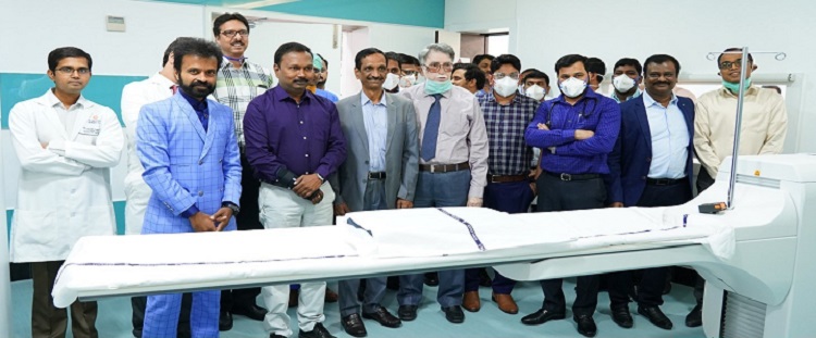
A state-of-the-art ‘Artificial Intelligence integrated PET-CT’ was inaugurated by Dr G Srinivas Rao, Director of Public Health & Family Welfare, Government of Telangana in the Nuclear Medicine Department at Yashoda Hospitals, Somajiguda.
“The new state-of-the-art artificial intelligence integrated PET-CT scanner at Yashoda Hospital Somajiguda is one more step towards our commitment to the early detection of cancer. The new scanner is now two times faster than the old generation scanners primarily due to the advanced technology known as ‘Time of Flight’. The scanner provides best quality images with reduced scanning duration and lesser radiation dose,” said Dr Rao.
Yashoda Hospitals Somajiguda is equipped with a comprehensive Nuclear Medicine set up providing services like PET-CT, Gamma camera imaging and radionuclide therapy under one roof. Apart from the newly upgraded imaging of FDG PET-CT, the department provides advanced and rare imaging like Ga-68 DOTA, Ga-68 PSMA, 18F DOPA PET-CTs, DAT imaging & WBC scans, apart from routine Gamma imaging like bone scan & renal scintigraphy.
“Yashoda Hospitals Somajiguda is one of the busiest and high volume centres of radionuclide therapies for thyroid cancer, neuroendocrine tumours, and prostate cancer. The centre also provides rare therapies like radiosynovectomy for inflammatory joint disease. Patients not only from Telangana and Andhra Pradesh but across India, visit us for these rare therapies. NextGen PET-CT is effective in the diagnosis of cancer, Endocrine Abnormalities and Neurodegenerative Disease,” said Dr Lingaiah Amidayala, Director – Medical Services, Yashoda Hospitals Group, Hyderabad.
The combined PET-CT Scan at Yashoda Hospitals, Somajiguda merges PET and CT images and provides detailed information about the size, shape and differentiating cancerous lesions from normal structures with accuracy. It is a diagnostic examination that combines two state-of-the-art imaging modalities and produces 3 dimensional (3D) images of the body based on the detection of radiation from the emission of positrons. It helps in the early detection of cancer and any potential health problem that reveals how the tissues and organs are functioning by identifying a variety of conditions.
Dr Hrushikesh Aurangabadkar and Dr A Naveen Kumar Reddy, Consultants in Nuclear Medicine while explaining about the PET-CT said, “The cancer cells require a great deal of sugar, or glucose, to have enough energy to grow. PET scanning utilises a radioactive molecule that is similar to glucose, called fluorodeoxyglucose (FDG). FDG accumulates within malignant cells because of their high rate of glucose metabolism. Once injected with this agent, the patient is imaged on the whole-body PET scanner to reveal cancer growth, which is usually difficult to characterize by conventional CT, X-Ray, or MRI.”